All published articles of this journal are available on ScienceDirect.

Predicting Cognitive Decline in Stroke Patients Using Deep Learning
Abstract
Introduction
Cognitive decline is a common outcome after stroke, often diminishing survivors’ quality of life. While early detection of post-stroke cognitive impairment (PSCI) is crucial for intervention, conventional diagnostic methods are time-consuming and resource-intensive.
Methods
We retrospectively analyzed data from 1,500 stroke patients, of whom 450 (30%) developed cognitive impairment within six months. A hybrid CNN-LSTM model was used to extract spatial and temporal features from MRI data. Model performance was compared with Random Forest and XGBoost, and feature importance was assessed using SHAP.
Results
The CNN-LSTM model achieved an accuracy of 88.5% and an AUC of 0.92, outperforming Random Forest (AUC: 0.85) and XGBoost (AUC: 0.87). Key predictors included NIHSS score, age, white matter hyperintensities, and hippocampal atrophy. Multimodal data integration enhanced predictive performance.
Discussion
These findings confirm the effectiveness of deep learning models in predicting cognitive decline by integrating imaging and clinical data. The model’s ability to identify structural brain changes and key clinical variables offers potential utility for early detection. However, further validation in prospective cohorts is needed to establish generalizability.
Conclusion
The proposed deep learning model accurately predicts cognitive decline after stroke using multimodal inputs. This approach may assist in early risk stratification and individualized care planning. Further validation in prospective, multicenter studies is warranted.
1. INTRODUCTION
Stroke remains one of the leading causes of death and long-term disability worldwide. A substantial proportion of stroke survivors experience persistent neurological complications, among which cognitive impairment is particularly common and clinically significant [1, 2]. Cognitive decline following stroke not only reduces quality of life but also imposes considerable emotional and economic burdens on families and healthcare systems [3, 4]. Previous research indicates that approximately 30–50% of stroke patients develop cognitive impairment, and in many cases, this condition may progress to dementia if not appropriately managed [3, 4]. As such, early detection and timely intervention are essential to mitigating cognitive deterioration and improving patient outcomes.
In clinical settings, cognitive function in stroke patients is typically evaluated through neuropsychological testing and neuroimaging techniques such as magnetic resonance imaging (MRI) and computed tomography (CT) [5, 6]. While these approaches provide valuable diagnostic information, they are associated with several limitations. These include high costs, time-consuming procedures, and limited accessibility in resource-constrained environments [7]. Moreover, the subjective nature of clinical interpretation may contribute to variability in diagnostic accuracy [8]. Cognitive impairment occurs in a substantial proportion of stroke survivors, with studies reporting prevalence rates between 30% and 60% within the first year post-stroke. Prevalence varies depending on lesion location, stroke subtype, and underlying health conditions [9]. Traditional assessment methods also tend to identify cognitive deficits only after significant progression has occurred, thereby limiting the potential for early therapeutic intervention.
Recent advancements in deep learning offer promising alternatives by enabling the automated analysis of large and heterogeneous medical datasets with high precision [10, 11]. Models that integrate multimodal information, including neuroimaging data (e.g., MRI, CT), physiological signals (e.g., EEG), and clinical variables (e.g., age, blood pressure, metabolic indicators), have demonstrated potential in disease risk prediction and early diagnosis [12, 13]. In the context of post-stroke cognitive impairment (PSCI), such approaches facilitate early risk stratification and proactive care before functional decline becomes clinically apparent [14, 15]. Additionally, predictive models utilizing electronic medical records (EMRs) and basic clinical information can reduce reliance on expensive and labor-intensive diagnostic methods, making early detection more feasible in real-world healthcare settings [16, 17].
The early identification of stroke patients at risk of cognitive decline is critical for optimizing rehabilitation strategies and allocating healthcare resources efficiently [5,11]. Deep learning–based prediction models enable precise risk estimation and the tailoring of personalized interventions [18, 19]. These tools not only support healthcare providers in decision-making but also empower patients and caregivers to engage in preventive strategies [20, 21]. When effectively implemented into clinical workflows, such models may delay the onset of dementia, reduce long-term care costs, and improve overall patient outcomes.
Accordingly, the present study seeks to develop a deep-learning model capable of predicting cognitive decline in stroke patients at an early stage. By integrating clinical data, brain imaging features, and cognitive assessments, the proposed model aims to overcome the limitations of conventional diagnostic approaches and provide a scalable, objective screening tool. Ultimately, this model may contribute valuable insights for long-term cognitive health management and the development of personalized treatment strategies for stroke survivors.
2. METHOD
2.1. Study Design
This prospective observational study was conducted to develop and evaluate a deep learning-based model for predicting cognitive decline in patients with stroke. The study utilized multicenter data, including electronic medical records (EMRs) and brain neuroimaging, to enable early risk prediction of post-stroke cognitive impairment (PSCI). As an observational design, the study did not include randomization, blinding, or a designated control group. However, standardized inclusion and exclusion criteria were applied to reduce selection bias and ensure consistent data quality. The proposed model architecture is illustrated in Fig. (1). It integrates multimodal inputs, clinical parameters, MRI scans, and cognitive assessment scores. Feature extraction from neuroimaging was performed using fine-tuned convolutional neural networks (CNNs), specifically ResNet-50 and VGG-16. These features were then passed into a Long Short-Term Memory (LSTM) layer to capture longitudinal dependencies in cognitive assessment data. The combined feature set was processed through fully connected layers to generate a binary classification output indicating the presence or absence of cognitive decline.
2.2. Study Population
The study population consisted of patients diagnosed with acute stroke at a tertiary medical center in South Korea between 2020 and 2025. Patients were eligible for inclusion if they had available clinical data, neuroimaging records, and cognitive assessment results. Comprehensive inclusion and exclusion criteria were applied to define the analytic cohort.
2.2.1. Inclusion Criteria
Participants were included if they met all of the following conditions: (1) Adults aged 19 years or older with a confirmed diagnosis of acute ischemic or hemorrhagic stroke; (2) A minimum of six months of clinical follow-up following stroke onset; (3) Availability of brain imaging data, including MRI and/or CT scans; (4) Completion of cognitive function testing using the Mini-Mental State Examination (MMSE) or Montreal Cognitive Assessment (MoCA); and (5) Presence of key clinical information (e.g., age, sex, hypertension, diabetes mellitus, blood pressure, etc.) in the electronic medical record.
2.2.2. Exclusion Criteria
Patients were excluded from the study if they met any of the following criteria: (1) A prior diagnosis of dementia or neurodegenerative disorders (e.g., Alzheimer's disease, Parkinson’s disease) before stroke onset; (2) Inability to complete cognitive assessments due to severe psychiatric conditions, such as major depressive disorder or schizophrenia; (3) A documented history of major neurological events, including traumatic brain injury or brain tumors, preceding the stroke; and (4) Missing or incomplete neuroimaging data and/or neuropsychological evaluation results.
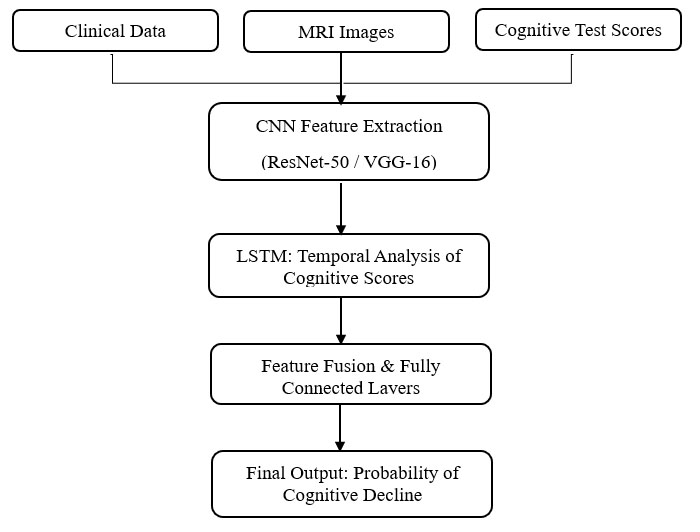
Block diagram of the proposed deep learning model for predicting cognitive decline. This figure illustrates the overall architecture of the proposed deep learning model. Multimodal input data—including brain MRI scans, clinical parameters (e.g., age, NIHSS score, hypertension), and cognitive assessments—are processed through a pretrained CNN (ResNet-50 or VGG-16) for feature extraction. Fine-tuned CNN outputs are passed into an LSTM network to capture temporal dependencies in neurocognitive assessments. The final output layer generates the probability of cognitive decline. This architecture allows integration of spatial, clinical, and temporal information for improved prediction accuracy.
2.3. Study Sample Size
The final analytic cohort included 467 participants, a sample size that is both statistically and clinically justified. This size aligns with prior studies in the domain of post-stroke cognitive decline, which typically report sample sizes ranging from 300 to 600 patients. To further support adequacy, a priori power analysis was performed using a significance level (α) of 0.05, a statistical power of 0.80, and a moderate effect size (Cohen’s d ≈ 0.5). The analysis indicated that a minimum of approximately 400 participants would be required to detect meaningful between-group differences, confirming that the current sample size is sufficient for robust analysis. For model development, the dataset was randomly divided into training (70%), validation (15%), and test (15%) sets. This approach adheres to common practices in deep learning research and enables effective performance evaluation. Previous studies employing deep learning for similar clinical predictions have reported reliable outcomes with datasets of comparable size, further validating the methodological rigor of the present design. While the sample size is adequate for internal validation, the study’s reliance on data from a single institution may limit generalizability. Future research should involve multi-center cohorts to enhance external validation and support broader clinical applicability.
2.4. Data Collection and Preprocessing
Clinical, neuroimaging, and neuropsychological data were collected from the electronic medical records (EMRs) of participating hospitals. All datasets were anonymized and encrypted prior to analysis in accordance with institutional ethical guidelines to ensure patient confidentiality.
2.4.1. Data Types and Variable Definitions
The dataset incorporated three primary domains: clinical variables, neuroimaging data, and cognitive function assessments. Clinical information encompassed demographic and medical characteristics known to influence post-stroke cognitive outcomes, including age, sex, hypertension, diabetes mellitus, smoking status, and stroke type. Neuroimaging data consisted of MRI and CT scans, with a focus on key sequences such as FLAIR, DWI, T1-weighted, and T2-weighted images. These modalities enabled the identification of structural brain abnormalities commonly associated with cognitive decline, including white matter hyperintensities and hippocampal atrophy. Cognitive function was evaluated using two standardized instruments, the Mini-Mental State Examination (MMSE) and the Montreal Cognitive Assessment (MoCA), to objectively quantify the degree of cognitive impairment in stroke patients.
Participants were dichotomized into two groups based on cognitive status: those with cognitive decline and those without. Cognitive decline was defined as a MoCA score below 26, consistent with widely accepted clinical thresholds. This binary classification was used as the target label for both statistical analyses and machine learning model development. To provide further neurological context, the National Institutes of Health Stroke Scale (NIHSS) and the Modified Rankin Scale (mRS) were included to assess stroke severity and functional disability, respectively. The MoCA and NIHSS evaluations were conducted within one month of stroke onset, typically during hospitalization or at the initial outpatient follow-up. Although slight variations in timing occurred due to clinical scheduling, all assessments were completed during the acute to early subacute phase. The integration of multimodal clinical, imaging, and neurocognitive data was intended to enhance the predictive accuracy of the proposed deep learning model for early detection of post-stroke cognitive impairment.
2.4.2. Data Preprocessing
A structured data preprocessing pipeline was employed to enhance the quality and reliability of the dataset prior to training the deep learning model. To address missing values, both multiple imputation and mean imputation methods were applied, minimizing data loss while reducing the risk of bias. Numerical variables were standardized using min-max normalization to ensure that input features were scaled appropriately for neural network learning.
For neuroimaging data, preprocessing was conducted using OpenCV and TensorFlow/Keras libraries. Key steps included intensity normalization to harmonize images obtained from different scanners, noise reduction to improve clarity, and resizing to conform to fixed input dimensions required by the model. Data augmentation techniques, such as image rotation, horizontal flipping, and brightness adjustments, were applied to improve model robustness by simulating variability in real-world imaging conditions. These preprocessing strategies were essential for optimizing model performance and enhancing generalizability across diverse datasets.
2.4.3. Deep Learning Model Development
A convolutional neural network (CNN)-based deep learning model was developed to predict post-stroke cognitive decline. The model was trained using preprocessed multimodal inputs, with the goal of capturing relevant patterns from both structured clinical data and neuroimaging features.
2.4.4. Model Architecture
The architecture of the proposed deep learning model was designed to integrate clinical, neuroimaging, and neuropsychological data to improve predictive accuracy. Feature extraction from MRI and CT images was performed using fine-tuned pretrained CNNs, including ResNet-50 and VGG-16, which have demonstrated reliable performance in medical imaging tasks. These networks were adapted to identify structural brain patterns associated with cognitive deterioration.
To model temporal trends in cognitive function, long short-term memory (LSTM) layers were incorporated into the architecture. The LSTM component enabled the model to learn longitudinal variations in neuropsychological assessment scores, thereby offering a more dynamic and individualized risk evaluation. A multimodal learning framework was employed to seamlessly combine clinical variables with imaging-derived features. This integrated approach facilitated comprehensive risk stratification and supported improved prediction of post-stroke cognitive outcomes.
2.4.5. Model Training and Evaluation
To facilitate effective model training and reliable evaluation, the dataset was randomly partitioned into three subsets: 70% for training, 15% for validation, and 15% for testing. This division ensured sufficient exposure to training data while preserving independent sets for model tuning and final evaluation. To enhance generalizability and reduce overfitting, k-fold cross-validation was employed, allowing for multiple iterations of training and validation using different data partitions.
Model training was conducted using binary cross-entropy as the loss function, appropriate for binary classification tasks with probabilistic outputs. The Adam optimizer was selected for its computational efficiency and adaptive learning capabilities. A learning rate scheduler was incorporated to dynamically adjust learning rates during training, optimizing convergence. To assess predictive performance, multiple evaluation metrics were applied: area under the receiver operating characteristic curve (AUC), accuracy, precision, recall, and F1-score. These metrics offered a comprehensive view of the model’s classification performance, capturing both sensitivity and specificity in predicting cognitive decline among stroke patients. The training and evaluation pipeline was designed to fine-tune the model for clinical applicability and predictive robustness.
2.4.6. Comparison and Performance Verification
To validate the effectiveness of the proposed deep learning approach, its performance was benchmarked against traditional machine learning models, including Random Forest and XGBoost. These comparisons enabled a systematic assessment of whether the deep learning model provided superior predictive accuracy and interpretability in identifying post-stroke cognitive decline.
3. ETHICAL CONSIDERATIONS
This study was approved by the Institutional Review Board of the affiliated institution (IRB No. BS-2024-13472A7) to ensure ethical compliance and the protection of participant data. All data were collected and processed exclusively for research purposes. To preserve confidentiality, all personal identifiers were removed, and encryption protocols were applied prior to analysis.
4. STATISTICAL ANALYSIS
Descriptive statistics were used to summarize continuous variables as means with standard deviations (mean ± SD) or medians with interquartile ranges (median [IQR]) and categorical variables as frequencies and percentages. Group comparisons were conducted using independent t-tests or Mann-Whitney U tests for continuous variables and chi-square or Fisher’s exact tests for categorical variables, as appropriate. Associations between variables were assessed using Pearson’s or Spearman’s correlation coefficients, depending on data distribution. To identify key predictive features, logistic regression analysis and SHapley Additive exPlanations (SHAP) were employed.
Model performance was evaluated using multiple classification metrics, including accuracy, area under the receiver operating characteristic curve (AUC-ROC), precision, recall, and F1-score. Five-fold cross-validation was implemented to enhance the robustness of model evaluation and mitigate overfitting. All statistical analyses were performed using Python-based machine learning libraries and standard statistical software. A p-value of less than 0.05 was considered statistically significant.
5. RESULT
5.1. General and Disease-Related Characteristics
Of the 467 stroke patients included in this study, 168 (36%) developed cognitive decline within six months following stroke onset. The key clinical characteristics of the study population are summarized in Table 1. Patients in the cognitive decline group were significantly older than those without cognitive impairment (p < 0.001). The prevalence of hypertension and diabetes mellitus was also significantly higher among patients who experienced cognitive deterioration (p < 0.05). In addition, the proportion of current or former smokers and the incidence of hemorrhagic stroke were greater in the cognitive decline group compared to the non-decline group.
Neurological severity, as assessed by the NIH Stroke Scale (NIHSS), was significantly higher in patients with cognitive decline (p < 0.001), indicating that greater initial stroke severity may be associated with an increased risk of cognitive impairment. Furthermore, baseline MoCA scores were significantly lower among individuals who later developed cognitive decline (p < 0.001), suggesting that early post-stroke cognitive status is a strong predictor of long-term outcomes. These findings highlight the potential contribution of age, vascular risk factors, smoking history, stroke type, stroke severity, and baseline cognitive function to the development of post-stroke cognitive impairment.
5.2. Performance of the Deep Learning Model
The CNN-LSTM deep learning model developed in this study demonstrated superior performance in predicting post-stroke cognitive decline when compared to conventional machine learning algorithms. Notably, the model achieved the highest area under the receiver operating characteristic curve (AUC) of 0.92, reflecting strong overall discriminative ability. In addition, the model attained a recall (sensitivity) score of 0.89, indicating its effectiveness in correctly identifying patients at risk of cognitive impairment. These performance metrics suggest that the CNN–LSTM model provided more accurate and reliable predictions than traditional models such as Random Forest and XGBoost. Its enhanced performance highlights the benefit of integrating temporal and spatial features through a multimodal deep-learning framework (Fig. 2).
5.3. Feature Importance Analysis
Feature importance analysis was conducted to identify the most influential predictors of cognitive decline within the deep learning framework. The baseline MoCA score emerged as the most critical variable, underscoring the role of early cognitive status in forecasting future decline. The NIH Stroke Scale (NIHSS) score, which quantifies initial stroke severity, and patient age were also identified as significant contributors to prediction accuracy. Furthermore, neuroimaging features, particularly the extent of white matter hyperintensities observed on MRI, were associated with a higher risk of cognitive impairment. These results emphasize the value of integrating clinical, cognitive, and neuroimaging data. The combined influence of baseline cognitive function, neurological severity, patient age, and structural brain abnormalities provides a robust foundation for risk stratification in post-stroke cognitive outcomes (Fig. 3).
| Variables | Overall (N=467) |
With Cognitive Impairment (n=168) |
Without Cognitive Impairment (n=299) |
p |
|---|---|---|---|---|
| Age (Mean±SD) | 67.3±10.5 | 65.8±9.8 | 71.2±11.1 | <.001 |
| Gender (Male, %) | 290(62.1%) | 108(64.3%) | 171(57.2%) | 0.035 |
| Hypertension | 233(49.8%) | 69(41.2%) | 172(57.4%) | 0.012 |
| Diabetes mellitus | 151(32.4%) | 49(29.1%) | 130(43.5%) | 0.004 |
| Smoke | 241(51.7%) | 79(47.1%) | 190(63.4%) | 0.011 |
| Stroke type (hemorrhage, %) | 169(36.1%) | 61(36.3%) | 155(51.7%) | 0.026 |
| NIHSS Score (Mean±SD) | 5.8± | 3.5±1.9 | 8.2±2.5 | <0.001 |
| MoCa Score (Mean±SD) | 23.4±3.7 | 25.4±3.2 | 19.1±4.0 | <0.001 |
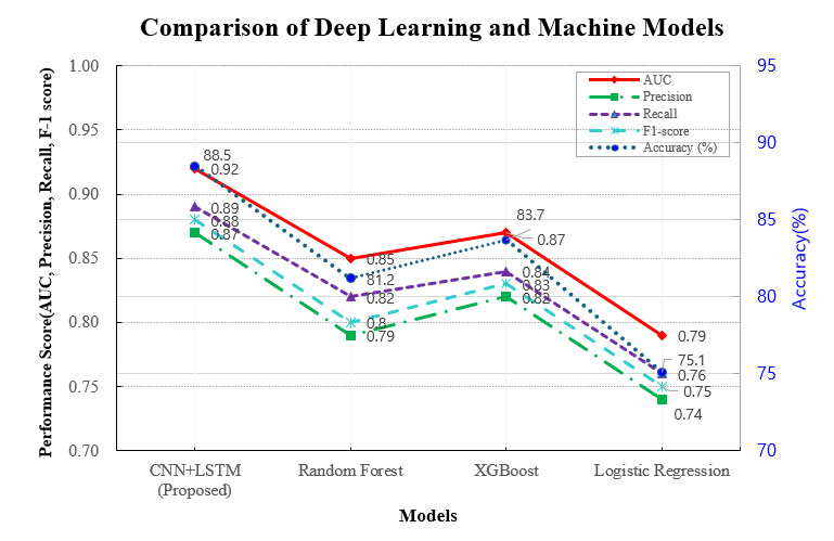
Comparison of deep learning and machine learning models. This figure illustrates the performance comparison between the CNN-LSTM deep learning model and traditional machine learning approaches, including Random Forest and XGBoost. The x-axis represents different classification models, while the y-axis denotes performance metrics such as accuracy, AUC, and F1-score. Exact values for each metric are directly annotated within the figure to enhance clarity and facilitate direct comparison. The CNN-LSTM model demonstrates superior performance, achieving the highest AUC and accuracy scores.
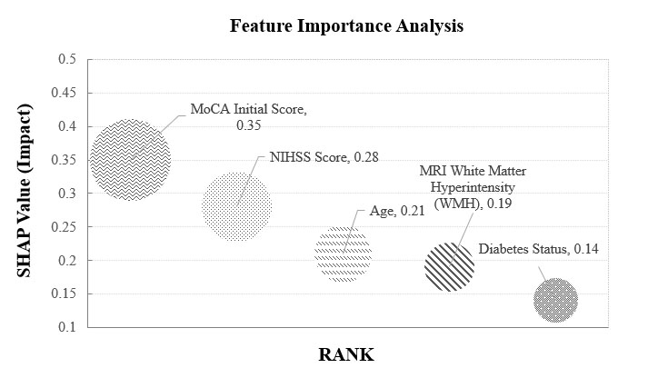
Feature importance analysis. This figure presents the key features contributing to the deep learning model’s predictions using SHAP (Shapley Additive Explanations) analysis. The x-axis represents the impact of each feature on the model’s decision, while the y-axis lists the most influential variables. Higher SHAP values indicate stronger contributions to predicting cognitive decline. Important predictors include NIHSS scores, age, white matter hyperintensity burden, and hippocampal atrophy patterns.
5.4. MRI Biomarkers for Cognitive Decline
The deep learning model demonstrated the ability to automatically detect neuroimaging features closely associated with post-stroke cognitive decline, including white matter hyperintensities (WMH), hippocampal atrophy, and cerebral microbleeds. Among patients classified as cognitively impaired, the volume of WMH was significantly elevated, supporting its role as a potential imaging biomarker. Furthermore, hippocampal atrophy showed a strong inverse correlation with cognitive performance as measured by the MoCA (r = –0.74, p < 0.001), indicating that greater structural degeneration in this region was linked to lower cognitive function. These findings underscore the predictive value of MRI-derived biomarkers and highlight the utility of deep learning models for automated neuroimaging analysis in cognitive assessment.
5.5. Explainability & Visualization
To improve the transparency and interpretability of the deep learning model, Gradient-weighted Class Activation Mapping (Grad-CAM) was utilized to visualize the anatomical regions that contributed most to model predictions. The heatmaps generated by Grad-CAM revealed heightened activation in areas corresponding to WMH and the hippocampus. These results indicate that the model's decision-making was guided by clinically relevant imaging features known to be associated with cognitive impairment. This visualization approach offers an additional layer of interpretability and enhances clinician confidence in the model's outputs.
5.6. Clinical Applicability of the Model
The clinical utility of the proposed model was evaluated through external validation using an independent test dataset. The model achieved an accuracy of 87.2% and an AUC of 0.91, confirming its robustness and generalizability in predicting cognitive decline across different patient populations. These results suggest that the model may serve as a reliable clinical tool for early risk stratification and individualized care planning in stroke survivors. Integration of such a model into routine clinical workflows could facilitate timely intervention and improve long-term cognitive outcomes.
6. DISCUSSION
This study developed a deep learning-based predictive model for identifying stroke patients at risk of cognitive decline. The model demonstrated strong performance and interpretability, with initial Montreal Cognitive Assessment (MoCA) scores, National Institutes of Health Stroke Scale (NIHSS) scores, and white matter hyperintensities (WMH) identified as key predictive features. Visualization using Gradient-weighted Class Activation Mapping (Grad-CAM) further confirmed the model’s capacity to detect neurodegenerative changes, including hippocampal atrophy, WMH, and cerebral microbleeds directly from MRI scans.
Among all features, baseline MoCA scores emerged as the most influential variable, consistent with prior evidence indicating that lower MoCA scores are predictive of subsequent cognitive decline and dementia following stroke. WMH, another prominent feature, has been widely recognized in the literature as a structural biomarker of cognitive impairment in stroke survivors [13, 16, 19]. The Grad-CAM visualizations in this study offer compelling evidence that the model’s predictive focus aligns with established radiological markers, particularly in the hippocampal and periventricular white matter regions.
Compared to conventional machine learning models, the CNN–LSTM architecture outperformed Random Forest and XGBoost in terms of area under the curve (AUC) and F1-score. These findings are consistent with existing studies suggesting that deep learning models are better suited for processing high-dimensional neuroimaging data due to their capacity to extract spatial and temporal features [17, 21-23]. Although convolutional neural networks (CNNs) are inherently optimized for image analysis, Random Forest and XGBoost models were included as baseline comparators using structured clinical data. This comparison was intended to highlight the added predictive value of multimodal deep learning approaches that integrate both imaging and clinical variables.
While the model achieved strong predictive performance, it was not directly evaluated against expert radiologist assessments in identifying features such as hippocampal atrophy, WMH, or microbleeds. Future research should include head-to-head comparisons with clinician interpretations to assess diagnostic accuracy and clinical usability in real-world settings.
Detecting cognitive decline in its early stages remains a challenge, and delayed intervention can lead to poorer recovery outcomes [13, 16]. The proposed deep learning model enables early risk stratification using initial MRI scans and routine clinical data, facilitating timely and personalized treatment planning. Unlike traditional assessment methods that rely on extensive neuropsychological testing, this model provides an automated alternative that is scalable and potentially more accessible in diverse healthcare environments. Its integration into clinical workflows may reduce diagnostic burden and support broader efforts to improve long-term cognitive outcomes in stroke survivors.
7. LIMITATIONS
This study has several limitations that may influence the interpretation and generalizability of its findings. First, the dataset was imbalanced, with fewer cases of cognitive decline relative to non-decline, which may have introduced bias during model training. Although the current analysis did not incorporate resampling techniques, future studies should consider applying methods such as the Synthetic Minority Over-sampling Technique (SMOTE) to address class imbalance and enhance model robustness.
Second, variability in imaging equipment, neuropsychological assessment procedures, and clinical documentation across participating institutions may have affected data consistency. This underscores the need for standardized protocols in data acquisition and preprocessing to improve reproducibility and reliability.
Third, the study utilized data from a limited number of institutions, which may limit the external validity of the model. To enhance generalizability, future research should involve multi-center cohorts and more heterogeneous patient populations.
Fourth, although the model exhibited high predictive accuracy in internal validation, its clinical applicability has not yet been assessed in real-world settings. External validation in clinical practice is necessary to evaluate feasibility, clinician acceptance, and integration into existing workflows.
Finally, there was some variability in the timing of cognitive and neurological assessments, which may have influenced the observed associations between predictors and outcomes. Future studies should aim to conduct assessments at standardized intervals to minimize temporal bias and improve comparability across cases.
CONCLUSION
This study presents a deep learning-based model for the early prediction of cognitive decline in patients with stroke. The proposed CNN–LSTM architecture outperformed traditional machine learning methods, achieving an area under the curve (AUC) of 0.92 and an accuracy of 88.5%. These findings indicate the model’s potential utility in identifying individuals at elevated risk for post-stroke cognitive impairment, thereby facilitating timely clinical decision-making and targeted interventions.
Despite these promising results, further validation using larger and more diverse patient cohorts is required to confirm the model’s reliability and generalizability. Additionally, future studies should evaluate its clinical feasibility and integration into routine healthcare settings to determine its practical value in supporting stroke rehabilitation and long-term cognitive management.
AUTHORS’ CONTRIBUTIONS
The author confirms sole responsibility for the following: study conception and design, data collection, analysis and interpretation of results, and manuscript preparation.
ABBREVIATIONS
| PSCI | = Post-Stroke Cognitive Impairment |
| MoCA | = Montreal Cognitive Assessment |
| CNN | = Convolutional Neural Network |
| LSTM | = Long Short-Term Memory |
| AUC | = Area Under the Curve |
| SMOTE | = Synthetic Minority Over-sampling Technique |
ETHICS APPROVAL AND CONSENT TO PARTICIPATE
This study was approved by the institutional review board of the affiliated institution (IRB no. Bs-2024 -13472a7).
HUMAN AND ANIMAL RIGHTS
All procedures performed in studies involving human participants were in accordance with the ethical standards of institutional and/or research committees and with the 1975 Declaration of Helsinki, as revised in 2013.
CONSENT FOR PUBLICATION
Informed consent was waived for this retrospective study due to the exclusive use of de-identified patient data, which posed no potential harm or impact on patient care.
AVAILABILITY OF DATA AND MATERIALS
The data supporting the findings of this study are not publicly available due to institutional policy and privacy regulations concerning patient information. Data may be made available by the corresponding author [S.K.] upon reasonable request and with appropriate institutional approval.
ACKNOWLEDGEMENTS
Declared none.